

How a common respiratory infection in an 8-month-old led to the diagnosis and correction of a rare birth defect by the pediatric team at NewYork-Presbyterian Brooklyn Methodist Hospital
By Amanda McCoy
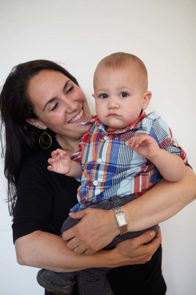
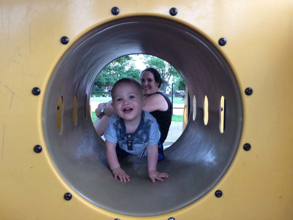
Nathan Rehm is a typical one year old. He loves to eat and seems to be growing bigger by the day. Looking at him today, it would be impossible to tell that only a handful of months ago he was diagnosed with a condition that threatened his life.
On an ordinary evening in late 2016, Joanne Spector and Alex Rehm were bathing their 8-month-old son when they noticed he appeared to be having difficulty breathing. Two days earlier, Nathan had been diagnosed with bronchiolitis, a relatively common respiratory infection in children, and the doctor told the parents to be on the lookout for certain abnormal signs that would require further medical attention. This was one of those signs.
The couple immediately took their son to their local urgent care facility, where they were referred to the ER, because the doctor there suspected that pneumonia might be the cause of Nathan’s symptoms. In the pediatric emergency room at NewYork-Presbyterian Brooklyn Methodist Hospital, a chest X-ray confirmed the pneumonia, but the doctors saw something else that was unrelated to the respiratory infection. The x-ray revealed an unusual mass on Nathan’s lung.
Spector had not been too worried about the bronchiolitis. An occupational therapist with the Helen Keller Services for the Blind Children’s Learning Center, she worked in a medical setting and knew that kids contracted respiratory infections all the time. But when the hospital asked to admit her son overnight, the worry began to set in.

“At that point, we were hoping it was just pneumonia,” said Spector. “We were admitted to the hospital, we got a room at one in the morning, and basically they were helping him with his breathing and doing a lot of suctioning and other medical treatments for the pneumonia It was scary, but the NYP Brooklyn Methodist staff let us put blankets on the floor (we had a little private room on the pediatric ward), and we brought some toys and made it as much fun as we could. Poor thing, he looked terrible, but we were able to keep his mood up and keep ours up by just taking it one step at a time.”
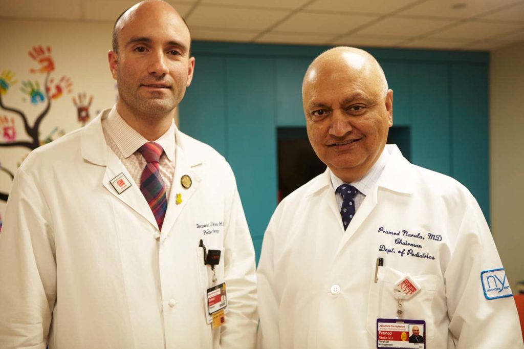
During their two-day hospital stay, the family met with Pramod Narula, M.D., a pediatric pulmonologist and Chairman of the Department of Pediatrics, who confirmed the presence of the suspicious mass and explained the need for additional testing. Because the young patient wasn’t showing any symptoms not typical of the pneumonia, he was sent home until he could fully recover from the infection.
Once the respiratory symptoms subsided, Dr. Narula, his associate, Kathy Garrett-Szymansky, along with Demetri J. Merianos, M.D., an attending pediatric surgeon at the hospital, were able to expedite scheduling and bring Nathan in for an ultrasound examination. After careful inspection, the physicians narrowed the cause down to one of two possibilities: diaphragm eventration (which wouldn’t warrant surgery) or a diaphragmatic hernia. Historically, doctors would have administered a CT scan to investigate the potential for these types of conditions, but according to Dr. Narula, a CT scan is the equivalent of 300 X-rays. “One of our goals is to avoid radiation in kids,” added the physician, who advised against the procedure. Instead, an MRI was recommended, which would involve anesthesia, but eliminate the child’s exposure to radiation.
“On December 22, we went for an MRI at NYP Brooklyn Methodist—under anesthesia, which is a big deal for a baby,” said Spector. “It gave us the diagnosis of a diaphragmatic hernia. That’s a hole in the diaphragm, so at some point in his life, either in utero or sometime afterwards, the contents of his abdomen had invaded his chest cavity…up near his lungs and his heart. Everything seemed to be in the wrong place.”
According to the doctors, congenital diaphragmatic hernias are very rare in children, occurring in less than .05% of births. They are separated into three categories: Bochdalek hernias, Morgagni hernias, and hiatal hernias. Of the three, 95% of reported cases fall in the Bochdalek subset, with hiatal hernias being the most rare. During his entire tenure at NYP Brooklyn Methodist, Dr. Narula had never before seen a case involving such a young patient. And unlike eventration of the diaphragm, a hiatal hernia in a child this age would require surgical repair.
“When you’re a few months old, the chance of this hernia not eventually causing problems is basically zero,” explained Dr. Merianos. “That hole is going to keep getting bigger, and more and more stuff will keep going up into the chest. It would have become harder to treat as he got older.”
The doctor explained that a normal chest cavity does have a small opening in the diaphragm for the esophagus to pass through, allowing food to travel from the mouth to the stomach. The diaphragm is supposed to fit snugly around the esophagus, but in Nathan’s case, the hole was much bigger than it should have been. In the past and still in many parts of the country, this type of procedure would require a large, open incision, resulting in a mandatory seven to 10 day recovery period in the hospital and a potentially nasty scar. Dr. Merianos had other plans for his patient.
“Minimally invasive surgery is less painful, reduces the chance of infection and problems with the wound, and leads to a much easier recovery. If an open procedure had been performed, the baby would have been in the hospital for at least a week and would have been in a lot more pain.”
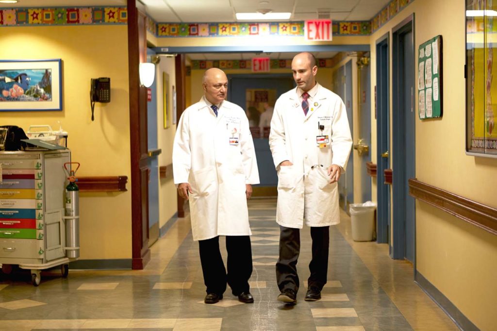
After graduating from New Jersey Medical School in 2004, the young doctor completed seven years of a residency in general surgery and a two-year fellowship in pediatric surgery before beginning his career as an attending surgeon at NYP Brooklyn Methodist Hospital. His extensive training and experience in minimally invasive surgery prepared him for a multitude of substantially less invasive operations, from a simple appendectomy to advanced laparoscopic and thoracoscopic procedures. So, rather than repair the young child’s diaphragm through a large cut, the surgeon was able to perform the entire procedure through four 5 mm laparoscopic incisions. No scarring, no lengthy recovery and substantially less pain.
“Minimally invasive surgery is less painful, more cosmetic, reduces the chance of infection and problems with the wound, and leads to a much easier recovery. If an open procedure had been performed, the baby would have been in the hospital for at least a week and would have been in a lot more pain,” added the doctor.
On January 25, the day of the surgery, three and half hours would pass before Nathan’s parents would see their son after kissing him goodbye in the pre-surgical prep room. Through only a few tiny incisions on the abdomen, Dr. Merianos and his team were effectively able to move every organ back where it belonged and tighten the hole with silk sutures. Although the surgeon initially planned on keeping his patient in the hospital for two full days while he monitored Nathan’s recovery, Dr. Merianos was pleased—surprised, even—to see the child respond very quickly. Nathan was able to go back to his Windsor Terrace home the next day.

“He’s doing fantastic,” added Spector of her son today. “He was coughing for a few weeks afterwards, and here I will give a shout out to Dr. Merianos again; even on the weekend, he was able to help us out when Nathan was coughing and we were afraid that something was wrong, and he was able to reassure us that it was a common side effect of the surgery. We had one follow-up appointment and that was it—the problem was solved. The whole team was terrific…NewYork- Presbyterian Brooklyn Methodist has just been a great hospital for us.”
NewYork-Presbyterian Brooklyn Methodist Hospital
506 6th Street, Brooklyn, NY 11215
718.780.3000 / nyp.org